Pages

management of breast symptoms


In any patient who present with breast lump or other symptoms suspicious of carcinoma, the diagnosis should be made by a combination of clinical assessment, radiological imaging and a tissue cytological or histological analysis the so called triple assessment.

starting with the clinical, lets imagine that a patient come to our clinic for the first time, so we take a proper history and do physical examination to reach our diagnosis. 

Then we can proceed with the imaging which are ultrasound and mammography but we must first know what are the indication for the patient. Ultrasound is suitable for women <35 years old because the breast tissue is dense and can distinguish cystic from solid lump. Compared to mammogram, it is for women >35 years old and routinely done in 2 views : cranio-caudal ( CC ) and mediolateral oblique view ( MLO ). 


ultrasound of breast


Mammogram.cranio-caudal view (CC) and the mediolateral oblique view, which is taken from an oblique or angled view


CC position


MLO position

 Next is pathology. We can do either fine needle aspiration or core cut needle ( Tru cut ) biopsy.Histology can be obtained under local anaesthesia using a spring-loaded core needle biopsy device. Cytology is obtained using a 21G or 23G needle and 10 ml syringe with multiple passess through the lump with negative pressure in the syringe. The aspirate is then smeared on to a slide, which is air dried or fixed. FNAC is least invasive technique of obtaining a cell diagnosis. 


CORECUT BIOPSY
FNAC


NG tube


 From it word, nasogastric tube means that this special tube is inserted to the nose until it reach the stomach. It is usually has different sizes . 

What is the indication of inserting nasogastric tube?

  • feeding for example in a case of dysphagia due to esophageal ca
  • gastric aspiration which we can use to analyze the pH of the stomach
  • stomach decompression in a case of intestinal obstruction
  • use in unconscious patient esp to prevent aspiration pneumonia
  • to make the bowel become inactive as the preparation for surgery

How long should we insert it? 
  • there is a scale on the tube which guide us to measure tubing : from the bridge of nose to the earlobe until to the point halfway between the end of the sternum and the navel


    STEPS

    Wear a gloves
    Patient should be in a sitting or 45 degree position. 

    Once we had note the distance of it, put the lubricant gel and insert the tube to the nose. Please check first is there any nasal obstruction so that we can avoid from put it from there because we are actually causing trauma to the nasal part. 
    Then we insert it to the posterior pharyngeal wall then continue until the patient will have a gag reflex . Ask the patient to swallow so that it would not enter the trachea. Finally u aspirate a sample of gastric contents. 

    Do not inject an air bolus, as the best practice is to test the pH of the aspirated contents to ensure that the contents are acidic. The pH should be below 6

    Secure tube with tape

    Look for colour,nature and amount of the gastric contents


    **In a unconscious patient, we use laryngoscope as an aid during the procedure and confirm the position of the tube by inject air and auscultate for air entry. Then do the x-ray.


    Complications
    • perforation of stomach
    • aspiration pneumonia
    • trauma
    • pain
    • loss of electrolytes
    • oesophagitis
    • necrosis 

      Burns

      salam. orait lets learn abour burns. heee since i had already learn it during 4th year and yesterday we have again a tutorial on it with Datuk ARA the plastic surgeon and surgery cluster head. okey lets go straight forward to our topic on burns. 

      Burn defined by any injury to skin by either thermal, chemical, electrical or radiation. There are high incidence of burns among low and moderate socioeconomic countries which the reasons might be due to :

      1. lack of education 
      2. overcrowding which cause house to house fire
      causes of burn??? yes as i have mentioned before. Then before we proceed, we should know a little bit about the anatomy and physiology our skin. 
      as we know, our skin has 3 layers. Epidermis, Dermis and Subcutaneous fat. Epidermis is the outermost layer and can be further subdivided .The second layer (located under the epidermis) is called the dermis; it contains nerve endings, sweat glands, oil glands, and hair follicles. Under these two skin layers is a fatty layer of subcutaneous tissue


      the simple example is sunburn. it can be categorized as superficial burns.


      FaMiLy plAnNiNg !

      hi everyone.assalamualaikum.
      okey just a short note on family planning.
      this is important for us to know there are many methods for a married couple to plan for their future/children
      and we should explain to them each and every methods have its own pros and cons and let them choose for their own benefit..we as a doctor can guide them to choose the best method


      case study 2

      Mrs BL, a 54 year old housewife, no known medical illness, presented to the clinic in Selayang Hospital with complaint of painless progressive enlargement of neck swelling since 5 years ago located at the center extending to the right side of the neck. It was associated with tiredness, insomnia, constipation and amenorrhea for 2 years. 


      INSPECTION
      The swelling moved with swallowing. The swelling is in the central part of the neck extending to the right side, ovoid in shape with the measurement of approximately 5cm in height, 10cm in length and 3cm in thickness. The skin overlying the swelling is smooth with no break and had no erythema. There are no dilated veins seen. There are no visible  pulsation seen.
      PALPATION
      On palpation, the swelling is well defined, smooth, soft and nontender. There was on nodule appreciated and it moves with swallowing. The isthmus and tracheal margin could not be appreciated. There were no palpable supraclavicular lymph node.
      PERCUSSION
      Percussion on the sternum was resonance.
      AUSCULTATION
      On auscultation, there was no carotid bruit heard.

      what's ur differential diagnosis???

      discovery PET-CT

      Assalamualaikum to all our muslim readers and good morning/afternoon/evening to everyone ^_^

      actually there are a lot of things that we can share together but because of my commitment to study so i can't post so much info/entry for this blog. maybe my partner/Dr can help you all.

      All right lets dont waste our time. i just want to share what I've learn from my radiology class yesterday. Actually to be honest, im not very good in this thing. ..plus i have to force myself to like this subject.hahaha... As a doctor, u should know this radiology stuff. U are responsible towards ur patient and u have to be familiar with it so that when ur consultant ask u to order like for example our topic today PET-CT for ur patient, at least u know what is PET-CT. 

      PET-CT
      PET : Positron Emission Tomography


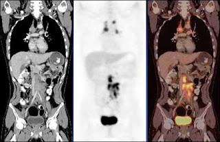
      If CT scan give u the information at the anatomical level, this PET-CT give u a picture at the cellular level before the organ distorted like for example the lymph node. Let say if someone have lymph node enlargement (due to many causes u just name it) but how can we know that the lymph node might change into cancer??? therefore by doing the PET-CT, it can show u .

      this PET-CT is actually give a radiation intravenously. A radiologist will inject a radiopharmaceutical 18-FDG/ Fludeoxyglucose  into the patient's vein. The fluorine-18 is combine with the glucose go to area where need a lot of glucose in the body such as cancer cells. As we know cancer cells need a lot of glucose to grow so it will absorb the 18-FDG and we can see on a picture like below that show the 18-FDG accumulate at the disease site.   


      " Using PET along side of CT can be beneficial because it provides an anatomical as well as a metabolic viewIt tells us what structure it is and what it is accomplishing biochemically. PET defines the cellular activity of the tumor, mass, etc. while CT provides details on the size and location of the tumor, mass, etc. "


      on ur left image : CT
      middle image: PET
      right image : PET-CT


      this is how the PET-CT machine looks like. Before we do the PET-CT, we should prepare the patient first. The normal thing that we ask the patient to do is fasting which start at 12 midnite till the next morning . 

      we should be aware of the patient condition because they should avoid strenuous exercise 24 hours before the scan done, should know also if any claustrophobic, diabetes, pregnant women,nursing mother because she should not breastfed her baby 24 hours after the scan done because we afraid of radiation transmission to the baby.

      The advantage of using PET-CT is that we can detect a disease earlier and accurately and the disadvantages of it are it is not easily available, expensive. In Malaysia there are only 3-4 PET-CT machine which 2 of it belongs to government : 1 in putrajaya hospital and another one in penang hospital. The other 2 belongs to private .  

      case study

      Mr Mok Lang, 67 year old Chinese gentleman, a known case of hypercholesterolaemia was admitted to Selayang Hospital with the chief complaint of right upper quadrant pain 5 days prior to admission. It is associated with yellowish discoloration of skin, vomiting and fever. He is an ex-smoker and occasional drinker.

      On examination, he is afebrile and other vital signs are normal. There is yellowish of sclera. There is guarding and tenderness over the right upper quadrant. There was hepatomegaly. Murphy’s sign positive.

      what's ur differential diagnosis????

      FACIAL NERVE LESION


      The picture above not relates to things that i want to share... But it seems like nice to see muslimah like this... if this are real. However these muslimah wearing purdah not bcoz of facial nerve lesion!!
      It is vital to know nerve relates to facial region especially to a doctor.. Face is an area that are very important for a dental surgeon. However , my blog partner don’t ever forget to review this matter and make a full review on full nerve lesion. As for my parts I try to revise nerve lesion relate to facial region only.


      How this can happen?


      SALIVARY GLAND DISEASE


      Salivary gland is one of the most wonderfull nikmat that’s Allah give to us. It is very important FOR eating, digestive system.. and so on.. then when we go older… we may notice our saliva become thicker because of the loss of serous secreting cells.. life is always like that.. nikmat that Allah give in this world are temporary  as compare to akhirat ..


      TRISMUS


      In dentistry , we doctors will have problem with this type of patients…. I said ‘ bukak, bukak lagi!!’ .. the patient said ‘huhuhu tak boleh la doctor!!’ … U know!! dont do this to ur patients!!   …. Hahahaha…   
      (*the things that I asked to open is their mouth / working area for a dental surgeon)…


      PIC 1.2 : INTRAORAL PIC/ CONGENITALLY MISSING TEETH


      PIC 1: OPG / CONGENITALLY MISSING TEETH


      Orthopantomogram shows Upper and lower arch , with normal general anatomical structures, No pathology seen on other anatomical structures, There were teeth germ missing for all 8’s upper and lower arches, 15, 35, 44, 45.
      11, 21 still erupting about 5mm intramucosally.
      Roots of 54, 64 already resorbed due to normal eruption of 14, 24.


      How to do EXAMINATION OF 'LUMPS AND BUMPS'


      LALALA…  what a nice morning.. wake up at 6 after did my subuh prayers.. thinking of something to do with LUMPS and BUMPS?? Hahaha… Do you know what should we observe / examine of lumps and bumps?? I try to list things in my mind… hahaha pretend to be clever!! Lets make things simple and sexy!!


      Copyright (c) 2010. Blogger templates by Bloggermint